
- Introduction
- CK5/6/TTF1
- Uses
Dual staining techniques have been used in immunohistochemistry. The use of these techniques is growing in many departments with a big advantage being that it can help in diagnosis where pathologist wish to distinguish between 2 different reactions.
Dual staining techniques, unlike single staining involves the use of two different stains. One of the most common procedures is to apply the Diaminobenzidinetetrahydorchloride (DAB) which produces a brown staining for the first or primary antigen then apply Alkaline phosphatase to stain up the secondary antigen of interest, as shown below.
Picture 1 – Dual staining use in Lung TTF1/CK5/6 staining
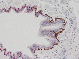
In the reaction shown below, the staining takes advantage of the ability of CK5/6 and TTF1 to stain up different cell populations. Using different processes of DAB staining for the TTF1 which is applied first producing the nuclear staining, while the use of alkaline phosphatase produces the red stain for CK5/6 positive areas. This as shown below allows a direct comparison between the two in the same tissue.
Picture 1 – Dual staining use in Lung TTF1/CK5/6
This is one of the most useful Dual staining techniques in ICC as it can be used to distinguish between adenocarcinoma and squamous cell carcinoma. However there are many, many applications for this porcess in immunocytochemistry and this is a area that is growing in importance.
Tumour or cell type |
CK 5/6 or TTF1 + or - |
Adenocarcinoma |
TTF1 + CK 5/6 - |
Squamous Cell Carcinoma |
TTF1 - Ck5/6 + |
Below is a table of some of the most common Dual staining techniques in immunocytochemistry, although this is changing daily in this expanding area for diagnosis.
Dual Stain |
Diagnostic use |
CK5/6/TTF1 |
Is very useful in distinguishing between adenocarcinoma and squamous cell carcinoma in lung biopsies. |
P16/Ki67 |
Testing using this is very effective in determination of whether cervical cancer is likely. In fact this has been shown to be more effective than screening using smears and maybe one reason for the decline in this area of cytology. http://www.ncbi.nlm.nih.gov/pubmed/24096620 |
P63/CK7 |
This staining is similar to CK5/6/TTF1 in helping distinguish between adenocarcinoma and squamous cell carcinoma and often used as an addition diagnostic test. It has limitations due to the poor specificity of CK7, which can be replaced by Napsin in some cases. |