
Immunohistochemistry - B Cells
- Introduction
- Bcl-2
- Cyclin D1
- CD20
- CD23
- CD79a
- Mum1
- Bob1
B cells belong to the group of white blood cells known as lymphocytes. B cells or B lymphocytes are a type of lymphocyte in the humoral immunity of the adaptive immune system. B cells can be distinguished from other lymphocytes, such as T cells by the presence of a protein on the B cell's outer surface known as a B cell receptor (BCR). This specialized receptor protein allows a B cell to bind to a specific antigen.
The principal functions of B cells are to make antibodies against antigens, to perform the role of antigen-presenting cells (APCs), and to develop into memory B cells after activation by antigen interaction. B cells also release cytokines (proteins), which are used for signaling immune regulatory function.
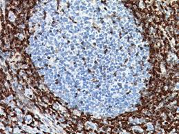
Bcl-2 is a gene that is used to encode for several proteins. Bcl-2 is very useful in immunohistochemistry as it is a very strong indicator of follicular B cell lymphoma and can distinguish this from other B cell lymphomas like Burkitts.
Bcl-2 shows a negative staining pattern in reactive geminal centers and positive staining pattern in neoplastic follicles in follicular lymphoma. Bcl-2 is also useful for diagnosis of cutaneous neoplams. The staining pattern of CD2 is cytoplasmic as shown below.
Picture 1 – Bcl-2 positivity in tonsil
Bcl-2 has several different applications. Below are a few examples.
Tumour or cell type |
Bcl-2 + or - |
Follicular B cell lympohoma |
Positive |
Basal cell carcinoma |
Positive |
Mantle cell lymphoma |
Positive |
Burkitt lymphoma |
Negative |
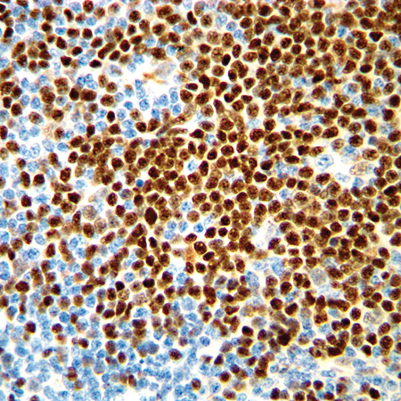
Cyclin D1 is a regulator in the cell G1 cycle. This is a very useful marker for determination of mantle cell lymphoma diagnosis. This is very useful in distinguishing between mantle cell lymphoma and other B cells tumours like burkitt or follicular lymphoma. As shown in the image below the staining pattern is nuclear.
Picture 2 – Cyclin D1 positivity in Mantle cell lymphoma
Cyclin D1 has several different applications. Below are a few examples.
Tumour |
Cyclin D1 + or - |
Mantle Cell lymphoma |
Positive |
Hairy cell leukaemia |
Some positive, very weak |
Diffuse large B cell lymphoma |
Positive |
Follicular Lymphoma |
Negative |
Burkitt cell lymphoma |
Negative |
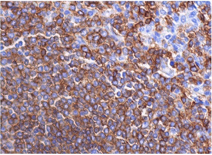
CD 20 or LC26 is a mature B cell marker. CD 20, although not as wide ranging as the Pan B cell marker as CD79a, is very useful for most mature B cell lymphomas. It is especially useful in combination with Cd79a to determine B cell orgins of tumours.
CD 20 is expressed on the membranes of B cells as shown in the image below.
Picture 3 – CD 20 positivity in Tonsil
CD 20 has several different applications. Below are a few examples.
Tumour or Cell type |
CD20 + or - |
All mature B cell tumours |
Positive |
Mature B cells |
Positive |
T cells |
Negative |
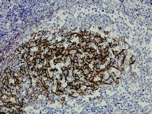
CD 23 is a useful B cell marker, useful to distinguish between tumours like Chronic lymphocytic leukaemia (CLL) and small lyphocytic lymphoma (SLL) from conditions like Mantle cell lymphoma.
As you can see below CD 23 has a membranous staining pattern.
Picture 4 – CD 23 positivity in Tonsil
CD 23 has several different applications. Below are a few examples.
Tumour |
CD23 + or - |
Chronic lymphocytic leukaemia |
Positive |
Small lyphocytic lymphoma |
Positive |
Mantle Cell Lymphoma |
Positive |
Burkit lymphoma |
Variable positive in about 76% of cases, but has specificity issues |
Hairy cell leukaemia |
Negative |
CD 79a is pan B cell marker, found in both immature and mature B cells, making it very useful in determination of tumour origin. This is found in B cell precursors but since it is sometimes lost on mature B cells it is often used with CD20 to confirm tumour origin on B cells. CD79a is also useful when treatment of a tumour has removed CD20 positivity as CD79a positivity is retained in these cases.
CD 79a has as shown below a membraneous staining pattern
Picture 5 – CD 79a positivity in Gastric adenocarcinoma
CD 79a has several different applications. Below are a few examples.
Tumour |
CD79a + or - |
All B cell lymphomas |
Positive |
All B cells |
Positive except in some mature B cells positivity has been lost |
Hodgkins Lymphoma |
Positive |
Plasma cell neoplasm |
Positive |
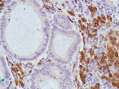
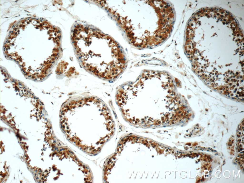
Mum1 or IRF-4 is found in plasma cells and also in small subset of geminal center B cells. It is also found in activated T cells as well. This is useful in combination with CD 30 in marking reed sternberg cells as well as in other lymphoid malignancy.
Mum1 has both a nuclear and cytoplasmic staining pattern as shown below.
Picture 6 – Mum1 positivity in Testis
Mum1 has several different applications. Below are a few examples.
Tumour or cell type |
Mum1 + or - |
Classic hodgkin lymphoma cells |
Positive |
Plasma cell neoplasm |
Positive |
Malignant Melanoma |
Positive |
Follicular B cell lymphoma |
Negative |
Burkitt Lymphoma |
Negative |
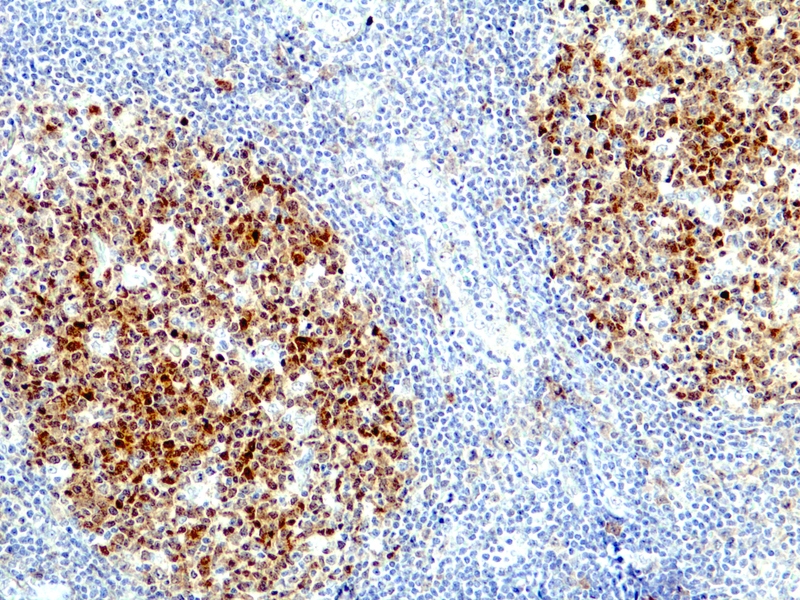
Bob.1 or OBF.1 is found on B lymphocytes, specifically in geminal center B cells, mantle B cells and plasma cells. This is very useful marker of cell different B cell lymphomas .
Bob.1 has a Nuclear staining pattern as shown below.
Picture 7 – Bob.1 positivity in Tonsil
Bob.1 has several different applications. Below are a few examples.
Tumour or cell type |
Bob.1 + or - |
Follicular B cell lymphoma |
Positive |
Mantle B cell Lymphoma |
Positive |
Diffuse large B cell lymphoma |
Positive |
Megakaryocytic acute myeloid leukaemia |
Negative |
Classic Hodgkin lymphoma |
Negative |